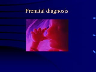
3. Introduction:
• Until the recent past, couples at high risk of genetic
disorder have the choose of:
- taking the risk
- considering other reproductive options (long term
contraception, sterilisation, termination of
pregnancy or even adoption and artificial
insemination (AID))
• until the 1966 when the relation of advanced
maternal age and increase rate of Down
syndrome was noticed and the prenatal
diagnosis was developed
4. • The purpose of prenatal
diagnosis is not simply to detect
abnormalities in fetal life and
allow termination. It rather have
the following goals:
5. • Provide a range of informed choice to the couples
at risk of having a child with abnormality
• Provide reassurance and reduce anxiety, especially
among high-risk groups
• Allow couples at high risk to know that the
presence or absence of the disorder could be
confirmed by testing
• Allow the couples the option of appropriate
management (psychological, pregnancy/delivery,
postnatal)
• To enable prenatal treatment of the affected foetus
6. Indications for prenatal diagnosis:
• advanced maternal age
• previous child with a
chromosome
abnormality
• family history of a
chromosome
abnormality
• family history of
single gene disorder
• family history of a
neural tube defect
• family history of other
congenital structural
abnormalities
• abnormalities
identified in pregnancy
• other high risk factors
(consanguinity, poor
obst., history, maternal
illnesses
7. Advanced maternal age
• Is the commonest indication for prenatal
diagnosis
• No standard criterion exists at what age women
should be investigated
• Most centres offer AMC or CVS to women aged
37 or over 35. (risk 1: 35)
• Figures differ for the risk of Down‘s sy. Because
a proportion of this pregnancy abort
spontaneously
8. Previous child with a chromosome abnormality
• Previous child with Down‘s sy due to non
disjunction or unbalanced translocation will give a
risk in subsequent pregnancy as, of mother‘s age
related risk plus 5%
• If one of parents have balanced chromosomal
rearrangement (translocation, inversion) causing a
serious problems for a previous child due to
unbalanced rearrangement, then recurrence risk is
between 1-2% and 15-20%. The risk will depend
on nature of rearrangement and segment involved
9. Family history of a chromosomal abnormality
• Usually no increase in risk compared to general
population since most chromosomal disorders will
arise as a result of disjunction than familial
rearrangement.
• A history of Down‘s sy
• However each situation should be confirmed by
nature of chromosome abnormality in affected
individual or urgent chromosome analysis from
blood of related parents if normal, no invasive
tests...
10. Family history of a single gene disorder
– A previous affected child
– Affection of one of the parents
– Positive family history
– Have a 25-50% recurrence and prenatal
diagnosis should be offered as many can be
diagnosed by DNA analysis or biochemical
testing (Achondroplasia, Huntington
disease, Neurofibromatosis,….)
11. Family history of a neural tube defect
• In first and second -degree relatives the risk
should be determined
• High risks were diagnosed by amniocentesis and
AFP assessment
• Ultrasound with MSAFP is the method of
diagnosis nowadays
• ? Small closed neural tube defects can be missed
even with the most skilled person (fortunately are
not associated with serious problems)
12. Family history of other congenital structural
abnormalities
• Evaluation of family pedigree
• Calculation of the risk
• If increased risk detailed ultrasound can
be offered between 16-18 weeks of pregnancy it
will detect most of serious defects
(cranial, cardiac, renal and limb deformation)
13. Abnormalities identified in pregnancy
• Uncertainty of maternal serum screening and fetal
anomaly scanning can make invasive procedure
for the diagnosis more necessary
• Poor fetal growth can be an indication for prenatal
chromosome analysis as well as for confirmation
of a serious and non-viable abnormality
14. Other high risk factors
• Parental consanguinity leading to hereditary
disorder or congenital anomalies (offer a detailed
ultrasound)
• Poor obst. history as recurrent miscarriage or still
birth indicating high risk in future preg. (offer
ultrasound of fetous and chromosome analysis of
parents)
• Maternal illnesses as poorly controlled DM or
maternal epilepsy treated with some drugs as
sodium valproate (offer detailed ultrasound)
17. Invasive methods of prenatal diagnosis
Amniocentesis
• Aspiration of 10-20 ml of amniotic fluid through
the abdominal wall under ultrasound guidance
around the 16 weeks of gestation.
• In about 14 days there will be enough cells for
chromosome analysis for biochemical or DNA
studies some time a longer time is needed for
grow of more cells.
• Couples should be informed of the risk of
abortions (0,5-1%) and the possibility of
termination if wished.
18. Chorionic villus sampling
• It enables diagnosis in first trimester (10-11 week
of gest.) under ultrasound guidance by transcervical
or transabdominal aspiration of chorionic villi
• These are fetal cells drived from the outer layer of
trophoblast.
• Results can be obtained in one to three days, so a
diagnosis in first trimester in addition that villi
provide a rich source of DNA
• Disadvantage is in higher risk of abortion (2-3%)
and limb abnormalities if carried before the 9
weeks of gestation.
19. Cordocentesis
• Visualisation of the umbilical vessels by
transabdominal ultrasound and enabling fetal
blood sampling.
• It is usually used in the management of Rhesus
isoimmunization and in some cases to solve the
problem of mozaicism.
21. Fetoscopy
• Visualisation of foetus by means of endoscope (it
has been suppressed by modern US
• It can be undertaken to diagnose a subtle structural
abnormalities pointing to a serious diagnosis
• Can also be used to obtain fetal samples for some
diagnosis as inherited skin disorders
(epidermolysis bullosa) and some metabolic
disorders in which enzymes are only in specific
organs
23. Non-invasive methods of prenatal diagnosis
Maternal serum AFP
• Mostly done around the16 weeks of gestation.
• More specific for the diagnosis of NTD (95% of
NTD can occur with out a history)
• Amniocentesis was used to confirm the diagnosis
but with a good detailed ultrasound first and
second degree can be diagnosed
• It has been found that by periconceptional
supplementation with folic acid decrease the rate
of occurrence of NTD and other abnormalities
24. Maternal screening test
• It is now a standard practice to offer screening for
NTD, Down‘s sy. and Edward sy. Using a blood
sample obtained from the mother at the 16 (15-20)
weeks of gestation
• It can diagnose up to 75% of NTD and 60-70% of
Down‘s sy.
25. Incresed
risk of
AFP UE3 HCG
Down’s
syn.
Dec. Dec. Inc.
Trisomy
18
Dec. Dec. Dec.
NTD Inc. Not
applicable
Not
applicable
26. Ultrasonography
• It offers a valuable means for prenatal diagnosis
• It is used for obst. diagnosis as placental
localisation and multiple preg. As well as for
prenatal diagnosis of structural abnormalities
which are not associated with known chromosome,
biochemical, or molecular defects.
• It is a non invasive with no risk to the foetus or
mother
• A specialised expensive equipment and a skilled
experienced operator are needed
27.
28. • It is offered to those with a history of genetic
disease
• Detailed fetal anomaly scanning is offered also to
all pregnant women around the 18 weeks of gest.
as a screening procedure for structural anomalies
(NTD and cardiac anomalies)
• It can identify features which suggest underlying
chromosomal abnormality indicating
amniocentesis.
29.
30. First Trimester US – Nasal Bone
Nasal bone present in a euploid
fetus in the first trimester
Nasal bone absent in a fetus with
T21in the first trimester
31. Second Trimester US – Nasal Bone
3D US coronal view in the second trimester
bilateral nasal bones present
32. Use of 3D US to improve the detection of absent nasal bone
3D/4D US
Benoit, 2005
41. Problems in prenatal diagnosis:
• Failure to obtain a sample or culture failure
• An ambiguous chromosome result
• An unexpected chromosome result
• Ultrasound soft markers
42. Prenatal treatment
• In the most situations the diagnosis of prenatal
abnormalities has a subsequent option of
termination of the pregnancy.
• While this applies in most situations, there is
cautious optimism that with the advent of gene
therapy prenatal diagnosis will, in time, lead to
effective treatment in utero.
43. Examples of gene therapy
• Treatment of the autosomal recessive
disorder - congenital adrenal hyperplasia
(CAH). Affected female are borne with
virilisation of the external genitalia. There is
an evidence that this can be prevented by
powerful steroid therapy at early gestational
age.
44. Gene therapy
Combined immunodeficiency
deficiency of the adenosine deaminase
bone marrow
retrovirus
Cystic fibrosis
deficiency of the transmembrane reg. gene
liposomes
fusing with epithelial cells
Haemophilia A
gene for factor VIII
liver tissue
application into portal vein
Lung carcinoma
K - ras (onkogene) at 30-40% adenocarcinomas
instillation of the mirror gene coding transfer of RNA
block of the decoding
p53 tum. suppressor gene at 50-70% of all carcinomas
instillation of good work. gene’s copy
retrovirus - into tumour deposit
45. • Treatment of a foetus affected with severe
combined immunodeficiency have been
reported. Transfused stem cells are
recognised as „self“ with the prospect of
good long term results. So immunological
tolerance of the foetus should make it easier
to commence such therapy before birth than
afterwards.
46. Summary of prenatal diagnosis (elements)
• It can be carried out by non-invasive procedures
(MS-AFP for NTD, triple test for Down‘s sy., and
US for structural abnormalities)
• Invasive procedures as amniocentesis or CVS is
usually requires for diagnosis of chromosome and
single gene disorders
• Invasive procedures convey small risk for
miscarriage (0.5-1% for amniocentesis, 2-3% for
CVS, and 3-5% for fetoscopy)
47. • The commonest indication of prenatal diagnosis is
advanced maternal age, family history of
chromosome single gene or structural abnormality
and a positive screening test in pregnancy
• While the significance of most prenatal diagnostic
findings is clear, situations can arise in which the
implications for the foetus are very difficult to
predict.
When this occurs the parents should be offered
specialised genetic counselling
48. Contingency Screening
First Step Second step % Pop
First trimester serum: have NT measured 40%
(PAPP-A, Free BHCG)
First trimester screen: have second trimester 20-25%
(PAPP-A, Free BHCG, NT) serum screening
First trimester screen: have first trimester 19%
(PAPP-A, Free BHCG, NT) targeted US (DV, TV,
Nasal Bone)
Very high risk 1:60 –
refer for CVS
Intermediate Risk –
Go to second step
Very low risk 1:1000 –
done
49. First trimester screening for
trisomy 21 by ultrasound and age*
*SP = screen positive rate 5%
Test Performance
NT T21 70-80%
NT + nasal bone T21 85-90%
Abnormal Ductus Venosus
waveform
T21 78% (at a SP rate of 1.7%
in a high risk population)8
NT + nasal bone +
abnormal DV
T21 >90% at a lower SP
rate 2-3%